Categories
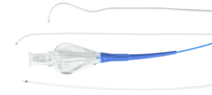
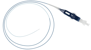
Procedure

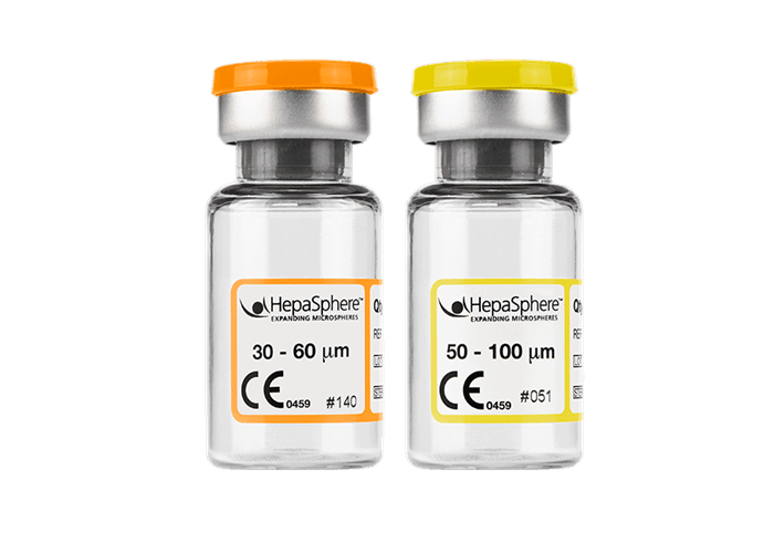
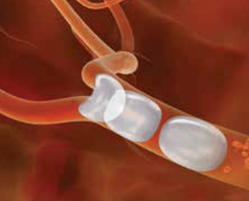 Illustration of the HepaSphere Microspheres conforming to the architecture of the vessel lumen
Once through the microcatheter, HepaSphere Microspheres rebound to their initial spherical shape with a consistent cross-sectional diameter.*
Affords atraumatic conformability to the architecture of the vessel lumen, providing contact surface area with the embolic material and the vessel intima, leading to a complete occlusion of the vessel.1,2
When in contact with non-ionic contrast media or normal saline (NaCI 0.9%) before delivery, HepaSphere Microspheres expand to approximately 4x their dry state diameter.* HepaSphere Microspheres are calibrated, spherical, hydrophilic microspheres made from 2 monomers (vinyl acetate and methyl acrylate) that combine to form a copolymer (sodium acrylate alcohol copolymer). This proprietary design allows a complete occlusion of the blood vessels.
HepaSphere Microspheres’ unique properties offer the following advantages:
Doxorubicin HCl used with HepaSphere Microspheres should be reconstituted with saline, never use pure water.
REFERENCES
1. De Luis et al. 2007. “In Vivo Evaluation of a New Embolic Spherical Particle (HepaSphere) in a Kidney Animal Model.” Cardiovasc Intervent Radiol 31, no. 2 (Mar–Apr): 367–76. doi: 10.1007/s00270-007-9240-1.
2. Bilbao et al. 2008. “Comparative Study of Four Different Spherical Embolic Particles in an Animal Model: A Morphologic and Histologic Evaluation. J Vasc Interv Radiol 19, no. 11 (Nov): 1625–38. doi: 10.1016/j.jvir.2008.07.014.
3. Gupta et al. 2011. “Hepatic Arterial Embolization with Doxorubicin-Loaded Superabsorbent Polymer Microspheres in a Rabbit Liver Tumor Model.” Cardiovasc Intervent Radiol 34, no. 5 (Oct): 1021–30. doi: 10.1007/s00270- 011-0154-6.
* Data on file.
409055001_001 ID 051425